


Camera reveals plants’ inner processes
Researchers take hundreds of plant pictures, exposing their biochemical activity
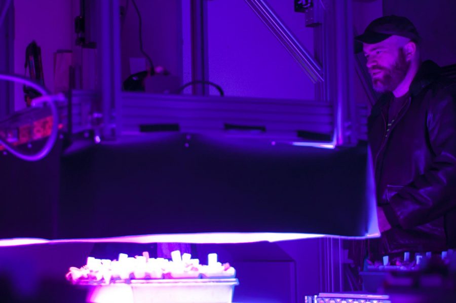
Magnus Wood scans plants with a Fluorescent imaging camera Wednesday at WSU’s Phonemics Center.
January 25, 2018
Scientists rapidly take pictures while flashing lights at plants, hoping to gain a better understanding of how they function.
WSU’s Phenomics Center, on the eastern edge of campus, operates an imaging camera system that can collect chlorophyll fluorescence data from plants when they are exposed to fluorescent light.
The camera, suspended close to the ceiling, navigates a metal grid above tables of plants.
Only 1 percent of the energy is emitted back into the camera. What the camera captures cannot be seen with the human eye, but it provides important diagnostic information for researchers. The plants appear different in pictures depending on their photosynthetic activity.
Helmut Kirchhoff, Phenomics Center coordinator and assistant professor for the Institute of Biological Chemistry, said the data can show genetic characteristics and how the plant creates energy.
He said the machine first operated in 2011, and that this is the easiest and least invasive way to monitor energy within plants. Kirchhoff estimated the machine could take between 200 and 300 pictures of plants simultaneously, which allows researchers to gather information efficiently.
“WSU tried to bring this phenomics approach further on campus,” Kirchhoff said. “There are different efforts going on now.”
Kirchhoff said the machine is computer-controlled and can program certain protocols, depending on what needs to be extracted from the data.
Magnus Wood, a scientific assistant at the Phenomics Center, explained how the machine functions step-by-step.
First, a protocol is set into the computer, which allows the machine to operate specific commands on its own, such as turning on at a given time or adjusting the length of the light pulses. Next, he chooses where to save the file and inputs coordinates to direct the camera.
The machine emits mostly red- and blue-colored light during screenings. These colors project rapid pulses that measure certain activities happening within the plant. The data researchers collect from the screenings can show them how the plant functions.
The camera within the machine is unable to pinpoint which plant is a mutant or non-mutant because it only develops a visual measurement of the plant. The researcher must to label which plant is what type in order to correctly interpret data.
Funding for the imaging camera system was through the Murdock Trust grant, which provides funds for universities within the Pacific Northwest. The proposal was made in 2015 and awarded on 2016.
Wood said there were plans for a similar imaging system to be built in Johnson Hall in the future.