
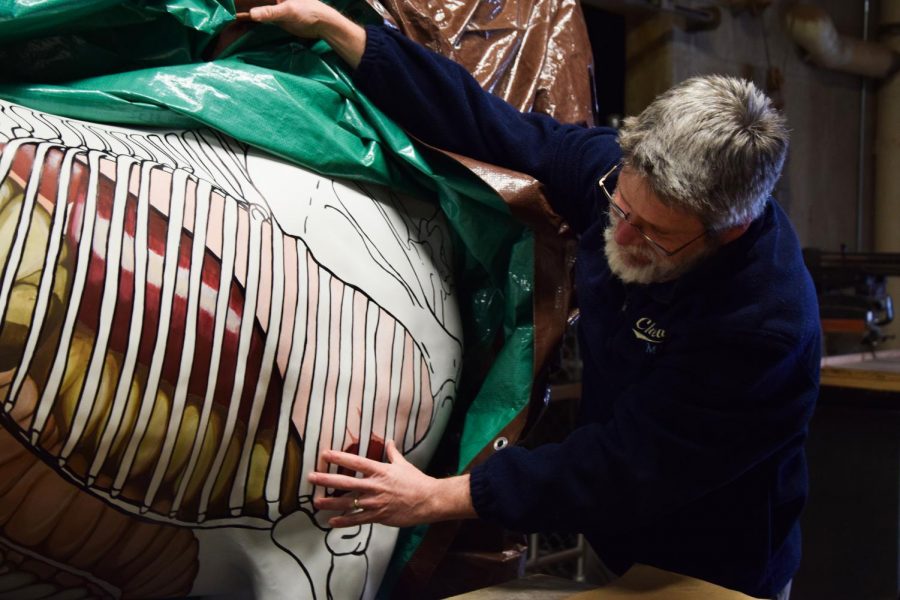
Painting horses for vet school courses
Graduate student puts final touch on donated glass animal for better classroom experience
JOSEPH GARDNER | THE DAILY EVERGREEN
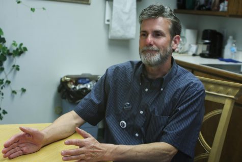
Patrick D. Wilson, clinical assistant professor in the Department of Integrative Physiology and Neuroscience, discusses the merits of using the painted life-size horse sculpture in a classroom setting Tuesday in McCoy Hall.
February 28, 2019
The fiberglass horse with organs painted on its thorax and abdomen may help students in the College of Veterinary Medicine learn about horse anatomy.
An anonymous donor gave $3,065 for the horse. The total cost, including shipping, was $2,125. Most of the money that remained went toward paying a student to paint it, Cynthia Faux, clinical assistant professor in the Department of Integrative Physiology and Neuroscience, wrote in an email.
Reanna Jarchow, a WSU student pursuing a doctorate of veterinary medicine, began to paint the horse last summer and has yet to finish it, Faux said. Jarchow still has to paint the vessels and nerves, like the jugular vein and arteries.
Patrick D. Wilson, clinical assistant professor in the Department of Integrative Physiology and Neuroscience, said he found and modified images of organs to project onto the horse so Jarchow could sketch and paint them on accurately. He included intestines, a liver, a spleen, a heart and lungs.
Faux said the visualization would help students better navigate the intricate placement of the animal’s organs.
“Putting the organs in a 3D relationship can be challenging to learn,” she said. “This is just a tool for students to be able to better map the anatomical topography for a live horse so they can visualize where the organs actually are.”
When students are doing physical exams, such as listening to a horse’s intestines with a stethoscope, they must know which section of a bowel they are listening to and where the other organs are in relation to their stethoscope, Faux said.
“[Students will] be looking at the real horse, and they need to visualize it, and they’ll go look at the fake horse,” Wilson said. “We encourage them also to go look at the animals that they’re dissecting and get that visual and then come back in and look at the live horse.”
Last semester, students did not get a chance to use the fake horse often, Faux said. The college will implement the horse in certain classes this semester. Some students will start using it as early as next week.
First-year students and students in the third-year applied anatomy class will use the model, but other groups can use it too, Wilson said.
It will be used for Horse Course, an annual one-day event used to educate people about horses, Faux said.
She said she came up with the idea of using a model horse with organs several years ago when she saw a different group using one.
“And I thought, ‘Wow, that’s pretty awesome, and I think that would be helpful for us,’” Faux said.